Leukemia/Lymphoma Phenotyping Evaluation by Flow Cytometry
Ordering Recommendation
Aids in the evaluation of hematopoietic neoplasms (ie, leukemia, lymphoma). Use to monitor therapy in patients with an established diagnosis of hematopoietic neoplasms.
New York DOH Approval Status
Specimen Required
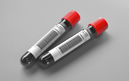
Bone marrow. Whole blood: Green (sodium heparin), lavender (K2EDTA), or pink (K2EDTA). Tissue or fluid.
Bone marrow: Transport 1 mL heparinized bone marrow (Min: 0.5 mL*)
Whole blood: Transport 5 mL whole blood. (Min: 1mL*)
Tissue: Transport 100 mg fresh tissue suspended in tissue culture media (e.g., RPMI 1640) (Min: 100 mg*)
Fluid: Transport 10-100 mL fresh fluid (Min: 3 mL*)
*Minimum volume is dependent on cellularity.
New York State Clients: Whole blood and bone marrow: Transport 2 mL (Min: 1 mL)
Tissue: Transport 0.2 cm3 suspended in RPMI.
Fluid: Transport 0.5 mL (Min: 0.5 mL) equal parts RPMI and specimen volume.
Do not send to ARUP Laboratories. Specimen must be received at performing laboratory within 48 hours of collection. For specimen requirements and direct submission instructions please contact ARUP Referral Testing at 800-242-2787 ext. 5161.
Specimen should be received within 24 hours of collection for optimal cell viability.
Bone marrow or whole blood: Room temperature. Also acceptable: Refrigerated.
Tissue or fluid: Refrigerated.
Clotted or hemolyzed specimens.
A minimum of 10,000 viable cells is required for flow cytometry phenotyping of samples containing a very limited number of markers (may also be called antibodies or antigens). For low-count specimens, supplying clinical and diagnostic information is especially important to help ensure the most appropriate marker combinations are evaluated before the specimen is depleted of cells.
Bone marrow or whole blood: Provide specimen source, CBC, Wright stained smear (if available), clinical history, differential diagnosis, and any relevant pathology reports.
Tissue or Fluid: Provide specimen source, clinical history, differential diagnosis, and any relevant pathology reports.
Follow up: If previous leukemia/lymphoma phenotyping was performed at another lab, the outside flow cytometry report and histograms (if possible) should accompany the specimen.
Ambient: 48 hours; Refrigerated: 48 hours; Frozen: Unacceptable
Methodology
Flow Cytometry
Performed
Sun-Sat
Reported
1-2 days
Reference Interval
Refer to report
Interpretive Data
Refer to report.
Analyte Specific Reagent (ASR)
Note
Flow cytometric immunophenotyping can aid in lineage assignment of acute leukemias and assist in diagnosis and classification of leukemias and lymphomas.
A panel of markers is performed on each case depending on the clinical history and specimen type. Without prior history or additional information, peripheral blood specimens typically undergo a preliminary analysis for B, T, NK, and myeloid/monocytic abnormalities, while bone marrows and lymph nodes undergo additional analysis for plasma cell abnormalities. Additional markers will be run as needed to further characterize abnormal populations.
Available Markers by population*:
T cell: CD1a, CD2, CD3, CD4, CD5, CD7, CD8, CD10, CD25, CD26, CD30, CD200, CD279 (PD-1), TCR gamma-delta, TRBC1, Cytoplasmic CD3
B cell: CD5, CD10, CD11c, CD19, CD20, CD22, CD23, CD25, CD38, CD103, CD123, CD200, surface Kappa, surface Lambda, cytoplasmic Kappa, cytoplasmic Lambda
Plasma cell: CD13, CD19, CD20, CD27, CD33, CD38, CD45, CD56, CD81, CD117, CD138, CD200, cytoplasmic Kappa, cytoplasmic Lambda
Myeloid: CD11b, CD13, CD14, CD15, CD16, CD33, CD34, CD36, CD38, CD41, CD42b, CD45, CD56, CD57, CD61, CD64, CD66b, CD71, CD117, CD123, HLA-DR, myeloperoxidase, glycophorin (CD235a), TdT
B lymphoblasts: CD10, CD19, CD20, CD22, CD24, CD38, CD45, CD58, CLRF2, cCD22, cCD79a
Mast cell: CD2, CD25, CD30, CD117, CD123
*Not all markers will be reported in all cases. Requests for specific markers to be run must be listed on manual requisition or by footnote for electronic orders. We do not offer individual marker identification separately outside of the markers in this panel.
The report will include a pathologist interpretation and a marker interpretation range corresponding to CPT codes of 2-8 markers, 9-15 markers, and 16+ markers interpreted. Charges apply per marker.
Hotline History
Hotline History
CPT Codes
88184, 88185 each additional marker; 88187 or 88188 or 88189.
Components
| Component Test Code* | Component Chart Name | LOINC |
|---|---|---|
| 0092286 | Number Of Markers | 19099-1 |
| 2008059 | Leuk/Lymph Phenotype, Impression | 45267-2 |
| 2013914 | Leuk/Lymph Phenotype, Source | 31208-2 |
Aliases
- Chronic Lymphocytic Leukemia Follow up Phenotyping by Flow Cytometry
- Follow-Up Phenotyping
- Hematopoietic neoplasms monitoring
- Leukemia/Lymphoma Evaluation Panel
- Leukemia/Lymphoma Phenotyping, Comprehensive - Bone Marrow
- Leukemia/Lymphoma Phenotyping, Comprehensive - Miscellaneous
- Leukemia/Lymphoma Phenotyping, Comprehensive - Whole Blood
